Maeda A, Kulbatski I, DaCosta RS; Mol Imaging. 2015 1;14:452-74
doi.org/10.2310/7290.2015.00022
Abstract
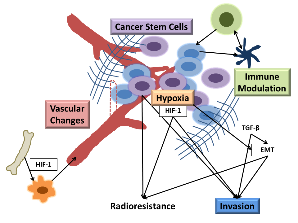
Radiation therapy is an effective cancer treatment used in over 50% of cancer patients. Preclinical research in radiobiology plays a major role in influencing the translation of radiotherapy-based treatment strategies into clinical practice. Studies have demonstrated that various components of tumors and their microenvironments, including vasculature, immune and stem cells, and stromal cells, can influence the response of solid tumors to radiation. Optically enabled imaging techniques used in experimental animal models of cancer offer a unique and powerful way to quantitatively track spatiotemporal changes in these tumor components in vivo at macro-, meso-, and microscopic resolutions following radiotherapy. In this review, we discuss the role of both well-established and emerging intravital microscopy techniques for studying tumors and their microenvironment in vivo, in response to irradiation. The development and application of new animal models, small animal microirradiation technologies, and multimodal optically enabled intravital microscopy techniques are emphasized within the framework of preclinical radiobiology research. We also comment on the potential influence that these newer imaging techniques may have on the clinical translation of new preclinical radiobiology discoveries.