Maeda A, Bu J, Chen J, Zheng G, DaCosta RS; Mol Imaging 2014;13(0):1-9
doi: 10.2310/7290.2014.00043
Abstract
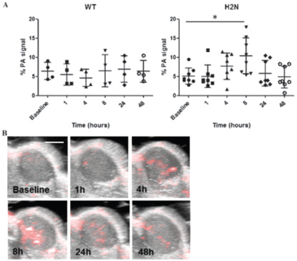
Biomarker-specific imaging probes offer ways to improve molecular diagnosis, intraoperative margin assessment, and tumor resection. Fluorescence and photoacoustic imaging probes are of particular interest for clinical applications because the combination enables deeper tissue penetration for tumor detection while maintaining imaging sensitivity compared to a single optical imaging modality. Here we describe the development of a human epidermal growth factor receptor 2 (HER2)-targeting imaging probe to visualize differential levels of HER2 expression in a breast cancer model. Specifically, we labeled trastuzumab with Black Hole Quencher 3 (BHQ3) and fluorescein for photoacoustic and fluorescence imaging of HER2 overexpression, respectively. The dual-labeled trastuzumab was tested for its ability to detect HER2 overexpression in vitro and in vivo. We demonstrated an over twofold increase in the signal intensity for HER2-overexpressing tumors in vivo, compared to low-HER2-expressing tumors, using photoacoustic imaging. Furthermore, we demonstrated the feasibility of detecting tumors and positive surgical margins by fluorescence imaging. These results suggest that multimodal HER2-specific imaging of breast cancer using the BHQ3-fluorescein trastuzumab enables molecular-level detection and surgical margin assessment of breast tumors in vivo. This technique may have future clinical impact for primary lesion detection, as well as intraoperative molecular-level surgical guidance in breast cancer.