2021

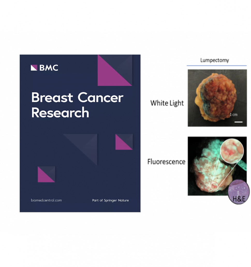
Intraoperative Fluorescence Imaging with Aminolevulinic Acid Decets Grossly Occult Breast Cancer: A Phase II Randomized Controlled Trial is published in Breast Cancer Research
970 1030 DaCosta Lab

After a lengthy journey, our paper “Intraoperative fluorescence imaging with aminolevulinic acid detects grossly occult breast cancer: a phase II randomized controlled trial” has been published in Breast Cancer Research as an open access article. The journal is widely read globally by general surgeons and breast cancer clinicians, so we are pleased it will receive broad clinical exposure and impact specifically in (breast) surgical oncology. This trial, led by Dr. DaCosta, was conducted at The Princess Margaret Cancer Center (Toronto, Canada) and demonstrates the importance of strong collaborations between our (imaging) scientists and clinicians at the Princess Margaret Cancer Center.  For…

read more
Intraoperative Fluorescence Imaging with Aminolevulinic Acid Detects Grossly Occult Breast Cancer: A Phase II Randomized Controlled Trial: Manuscript accepted to Breast Cancer Research
1030 606 DaCosta Lab

In 2009, the first patient was enrolled at Princess Margaret Cancer Center in this Phase II randomized controlled trial led by Dr. DaCosta in collaboration with surgeons Drs. Wey Leong and Alexandra Easson and pathologist Dr. Susan Done. The goal was to evaluate the clinical safety, feasibility and diagnostic performance of the PRODIGI handheld fluorescence imaging device combined with 5-ALA HCl for intraoperative visualization of invasive breast carcinomas. The Phase II trial was supported in large part by a $3M CIHR grant awarded to the DaCosta lab. Today, over a decade later, the manuscript for this study was accepted for…

read more
In Vivo Mesenchymal Progenitor Cell Dynamics: Dr. Niloufar Khosravi publishes in Biomaterials
818 807 DaCosta Lab

Niloufar Khosravi, a DaCosta Lab PhD student alumna, published in Biomaterials today: “New insights into spatio-temporal dynamics of mesenchymal progenitor cell ingress during peri implant wound healing: Provided by intravital imaging”. Abstract Surface topography drives the success of orthopedic and dental implants placed in bone, by directing the biology occurring at the tissue-implant interface. Over the last few decades, striking advancements have been made in the development of novel implant surfaces that enhance bone anchorage to their surfaces through contact osteogenesis: the combination of the two phenomena of recruitment and migration of mesenchymal progenitor cells to the implant surface, and…

read more
Privacy Preferences

When you visit our website, it may store information through your browser from specific services, usually in the form of cookies. Here you can change your Privacy preferences. It is worth noting that blocking some types of cookies may impact your experience on our website and the services we are able to offer.

Click to enable/disable Google Analytics tracking code.
Click to enable/disable Google Fonts.
Click to enable/disable Google Maps.
Click to enable/disable video embeds.
Our website uses cookies, mainly from 3rd party services. Define your Privacy Preferences and/or agree to our use of cookies.