
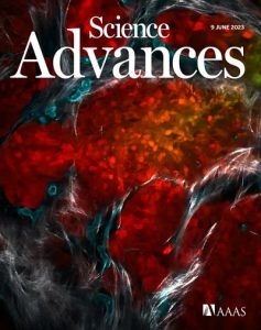
Our recent publication, “Quantitative intravital imaging for real-time monitoring of pancreatic tumor cell hypoxia and stroma in an orthotopic mouse model,” which was selected for the cover of the journal Science Advances, was announced on the UHN Research News portal.
“A research team led by Dr. Ralph DaCosta, Senior Scientist at the Princess Margaret Cancer Centre, recently developed an innovative optical imaging platform that can monitor pancreatic tumours in real time and at the cellular level over prolonged periods in experimental models of human pancreatic cancers.
The technique is shedding new light on the tissues and blood vessels that surround pancreatic tumour cells as they grow—known as the tumour microenvironment. This tumour microenvironment is characterized by low oxygen (called hypoxia) and excessive fibrous or connective tissue that can have a significant impact on the effectiveness of anticancer treatments.
“While understanding the microenvironment is key to developing better ways to treat the disease, studying it has been a challenge. This is due to its complexity and the fact that the composition of the microenvironment is constantly changing as tumour cells grow and spread into nearby tissues, all the while modifying the environment in which they are growing,” says Dr. DaCosta.
To address this, the researchers developed a method called quantitative intravital microscopy, a type of imaging that enables researchers to visualize pancreatic tumour progression, measure oxygen levels within tumour cells and observe changes in the supportive tissues and blood vessels surrounding the tumour.
Using fluorescence-based probes, the team tracked cell oxygen levels in the tumour and in surrounding tissues over time for several weeks in an experimental model. Their findings revealed spatial variations in the structure and function of blood vessels within the tumour microenvironment and in oxygen levels within tumours.
“Understanding the interplay between oxygen levels in tumour cells and those of the nearby microenvironment is crucial for developing effective treatment strategies—especially when oxygen levels fall and the tissues become hypoxic,” explains Dr. DaCosta. “Our novel imaging platform provides valuable insights into the dynamic nature of hypoxia within the pancreatic tumour microenvironment, offering potential targets for therapeutic interventions.”
While further research is needed to fully understand the complexities of the pancreatic tumour microenvironment, this study represents a significant step forward in unraveling the mysteries surrounding pancreatic cancer and provides hope for the development of more effective treatment options.
The paper was selected for the cover of the journal Science Advances, illustrated below.
This work was supported by the Terry Fox Research Institute and The Princess Margaret Cancer Foundation.
Samuel T, Rapic S, O’Brien C, Edson M, Zhong Y, DaCosta RS. Quantitative intravital imaging for real-time monitoring of pancreatic tumor cell hypoxia and stroma in an orthotopic mouse model. Sci Adv. 2023 Jun 9;9(23):eade8672. doi: 10.1126/sciadv.ade8672.
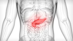
The pancreas is an organ within the abdomen that plays a major role the body’s digestive system via the secretion of enzymes and hormones. Pancreatic cancer is a deadly disease and the third leading cause of cancer-related death in Canada. Only 10% of those diagnosed with the condition survive beyond five years.”
Please see the announcement here.