We are excited to announce that our paper was chosen for the cover of Science Advances!
The DaCosta Lab is pleased to announce that our recent publication, “Quantitative Intravital Imaging for Real-time Monitoring of Pancreatic Tumor Cell Hypoxia and Stroma in an Orthotopic Mouse Model” was chosen to be on the cover of Science Advances.
Link to publication: https://www.science.org/doi/10.1126/sciadv.ade8672
Authors: Dr. Timothy Samuel, Dr. Sara Rapic, Cristiana O’Brien, Michael Edson, Yuan Zong, and Dr. Ralph S. DaCosta
Abstract: Pancreatic cancer is a lethal disease with few successful treatment options. Recent evidence demonstrates that tumor hypoxia promotes pancreatic tumor invasion, metastasis, and therapy resistance. However, little is known about the complex relationship between hypoxia and the pancreatic tumor microenvironment (TME). In this study, we developed a novel intravital fluorescence microscopy platform with an orthotopic mouse model of pancreatic cancer to study tumor cell hypoxia within the TME in vivo, at cellular resolution, over time. Using a fluorescent BxPC3-DsRed tumor cell line with a hypoxia-response element (HRE)/green fluorescent protein (GFP) reporter, we showed that HRE/GFP is a reliable biomarker of pancreatic tumor hypoxia, responding dynamically and reversibly to changing oxygen concentrations within the TME. We also characterized the spatial relationships between tumor hypoxia, microvasculature, and tumor-associated collagen structures using in vivo second harmonic generation microscopy. This quantitative multimodal imaging platform enables the unprecedented study of hypoxia within the pancreatic TME in vivo.
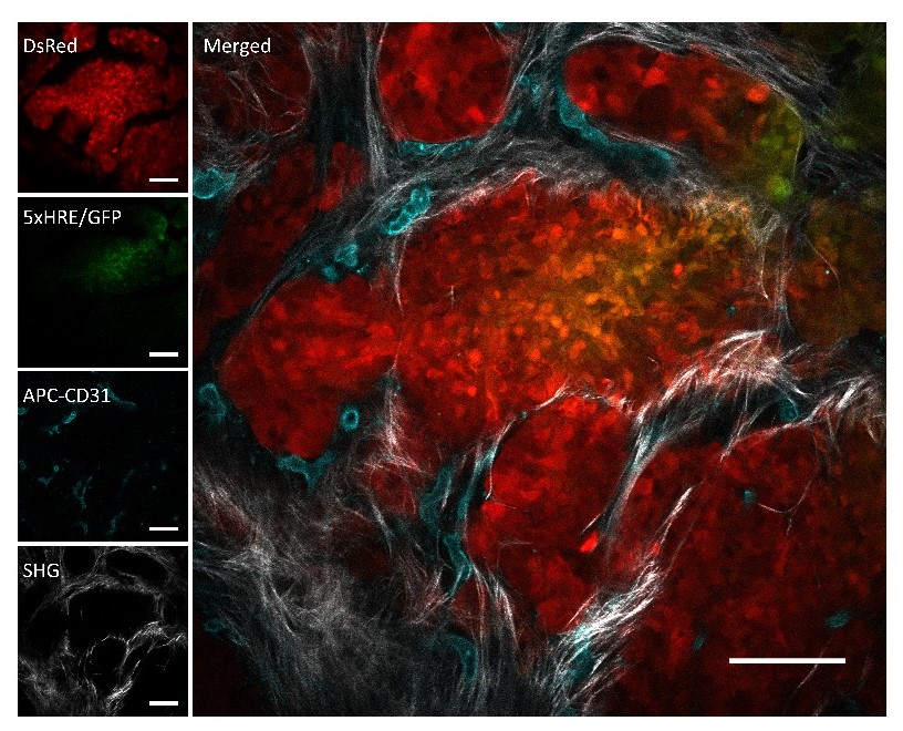
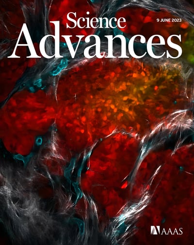
A representative image of an in vivo orthotopic BxPC3-DsRed-5xHRE/GFP tumor foci, taken using intravital fluorescence and SHG microscopy.
BxPC3 cells (DsRed) are shown in red, HIF activity (5xHRE/GFP) in green, blood vessels (APC-CD31) in cyan, and fibrillar collagen (SHG) in white. Scale bars, 100 μm