Chamma E, Qiu J, Lindvere-Teene L, Blackmore KM, Majeed S, Weersink R, Dickie CI, Griffin AM, Wunder JS, Ferguson PC, DaCosta RS; J Biomed Opt. 2015 Jul 1;20(7):76011
doi: 10.1117/1.JBO.20.7.076011.
Abstract
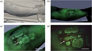
Standard clinical management of extremity soft tissue sarcomas includes surgery with radiation therapy. Wound complications (WCs) arising from treatment may occur due to bacterial infection and tissue breakdown. The ability to detect changes in these parameters during treatment may lead to earlier interventions that mitigate WCs. We describe the use of a new system composed of an autofluorescence imaging device and an optical three-dimensional tracking system to detect and coregister the presence of bacteria with radiation doses. The imaging device visualized erythema using white light and detected bacterial autofluorescence using 405-nm excitation light. Its position was tracked relative to the patient using IR reflective spheres and registration to the computed tomography coordinates. Image coregistration software was developed to spatially overlay radiation treatment plans and dose distributions on the white light and autofluorescence images of the surgical site. We describe the technology, its use in the operating room, and standard operating procedures, as well as demonstrate technical feasibility and safety intraoperatively. This new clinical tool may help identify patients at greater risk of developing WCs and investigate correlations between radiation dose, skin response, and changes in bacterial load as biomarkers associated with WCs.