We would like to welcome a new research technician to our lab:

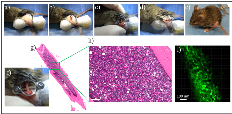
Emily was previously part of our lab from 2009-2016, during which she helped to pioneer our first animal window chamber models to study the tumor microenvironment. In re-joining our lab, Emily will be responsible for conducting animal surgeries involving the use of our novel window chamber systems for direct microscopy imaging of human orthotopic tumors. Emily will also help lead the development and validation of new intravital imaging models of cancers including solid cancers (e.g. pancreatic, brain and breast tumors) and hematological cancers (e.g. leukemia). Our lab pioneered the use of intravital multimodal imaging of cancers to study treatment response specifically within the tumor microenvironment; this has led to important new discoveries through local, national and international collaborations.

Welcome back Emily, we are excited that you will help to expand our cancer imaging research program by developing new animal cancer models that will enable new cancer discoveries at the Princess Margaret Cancer Centre!
