Investigating the Effects of Stereotactic Body Radiation Therapy on Pancreatic Tumor Hypoxia and Microvasculature in an Orthotopic Mouse Model using Intravital Fluorescence Microscopy
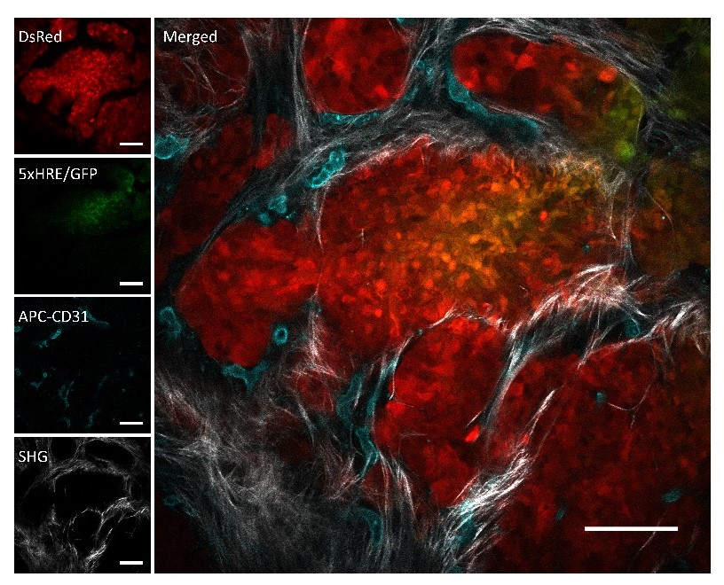
https://dacostalab.ca/wp-content/uploads/2025/01/Sci-Reports-Nature.jpg 1000 625 DaCosta Lab DaCosta Lab https://dacostalab.ca/wp-content/uploads/2025/01/Sci-Reports-Nature.jpgOur recent paper, “Investigating the Effects of Stereotactic Body Radiation Therapy on Pancreatic Tumor Hypoxia and Microvasculature in an Orthotopic Mouse Model using Intravital Fluorescence Microscopy,” was published in Scientific Reports (December 2024). Background and Aims: Despite decades of improvements in cytotoxic therapy, the current standard of care for locally advanced pancreatic cancer (LAPC) provides, on average, only a few months of survival benefit. Stereotactic Body Radiation Therapy (SBRT), a technique that accurately delivers high doses of radiation to tumors in fewer fractions, has emerged as a promising therapy to improve local control of LAPC; however, its effects on the tumor microenvironment and hypoxia remain poorly understood. To explore how SBRT affects pancreatic tumors, we combined an orthotopic mouse model of pancreatic cancer with an intravital microscopy platform to visualize changes to the in vivo tumor microenvironment in real-time. Methods: Mice received SBRT (5 × 8 Gy) or were left untreated, and were imaged before and 1, 4, 7, and 14 days after treatment (n = 7/group). A fluorescent human pancreatic cancer cell line (BxPC3-DsRed) engineered to express GFP under hypoxic conditions (driven by hypoxiainducible factor, HIF) was used to monitor tumor hypoxia. Immunohistochemical staining was also performed on tissues to validate in vivo data. Results: Our findings demonstrate a persistent decrease in pancreatic tumor hypoxia as early as one day after SBRT. This coincided with a decrease in both tumor cell proliferation and cell density in the SBRT group. Reduced demand for oxygen after SBRT (due to cell death and growth arrest from treatment) significantly contributed to reoxygenation of the pancreatic TME. Conclusions: Understanding how this reoxygenation phenomenon occurs in a dose-dependent manner will help improve dosing and fractionation schemes for clinical SBRT.
read more